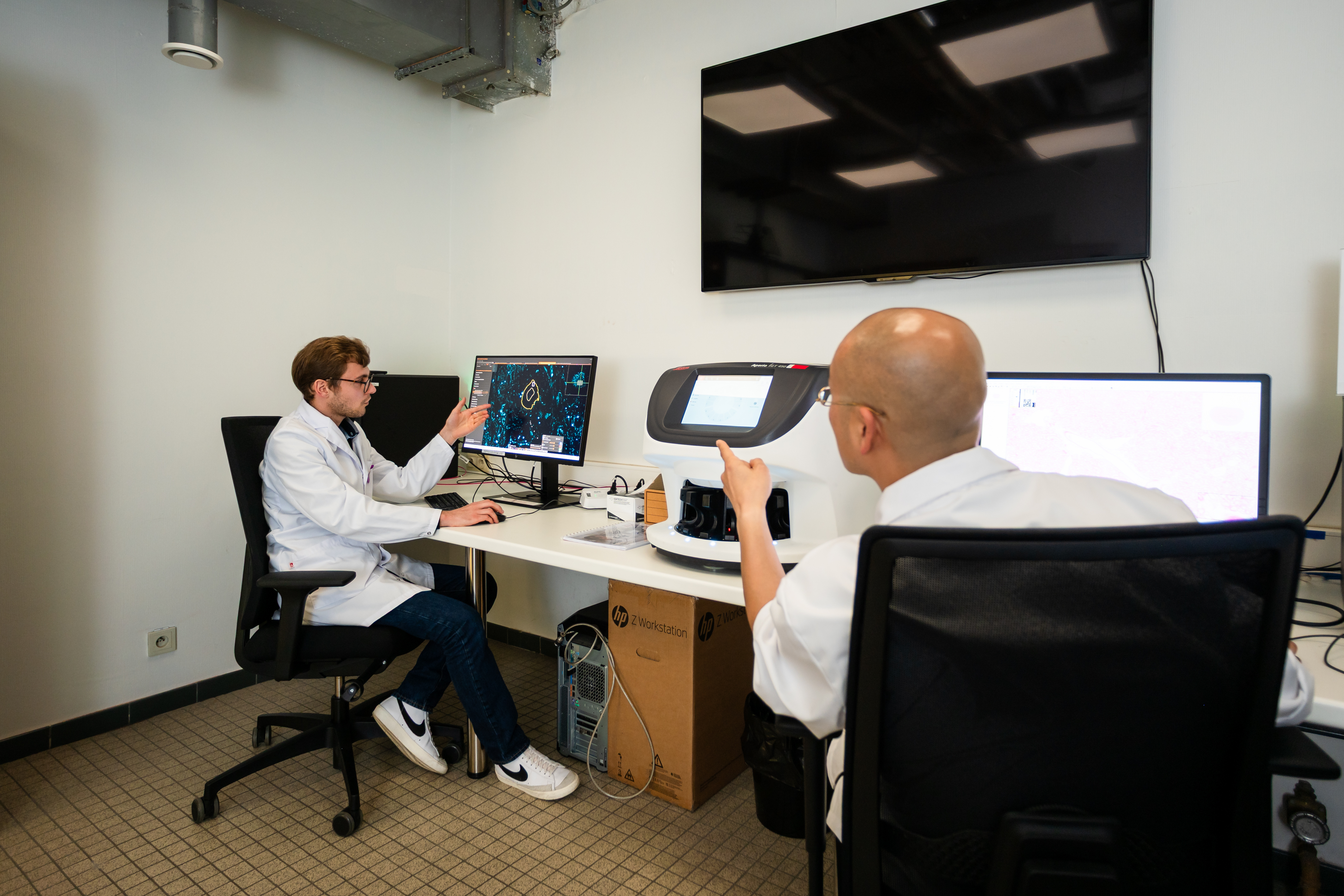
“Looking through a microscope is no longer necessary. The Comet instantly delivers digital images.”


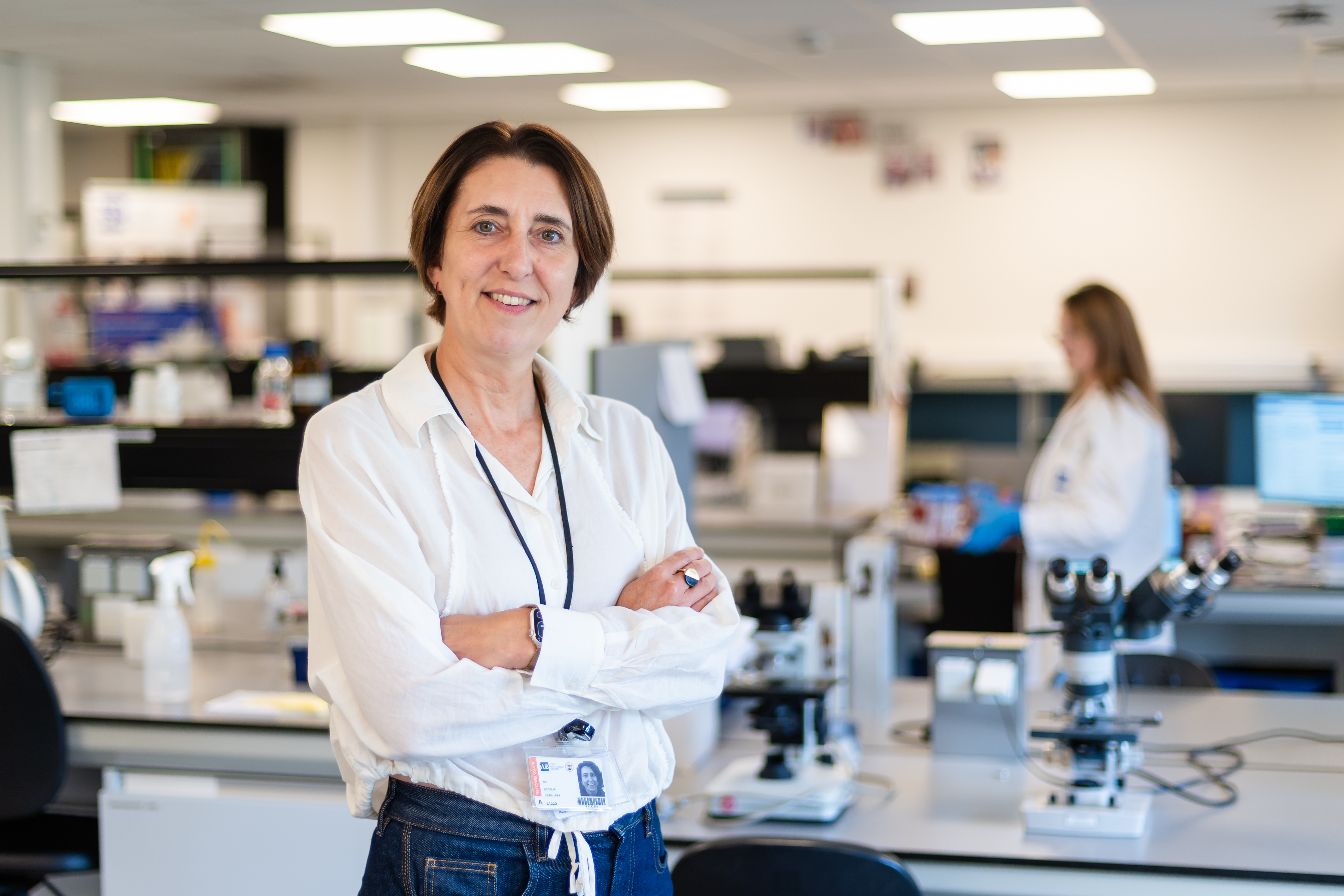
The Visual and Spatial Tissue Analysis (VSTA) research centre is equipped with advanced techniques that reveal where specific genes or proteins are active within a tissue sample. As a core facility, VSTA welcomes researchers both from within and beyond VUB, explains VSTA director Professor Ilse Rooman. “We’ve just purchased a new device, the Comet. If our researchers want to remain competitive, we have no choice but to invest in costly technology.
VSTA is located on the ground floor of Block D at the UZ Brussel campus in Jette. During our visit, the much-anticipated device is not yet visible. In the corridor, Ilse Rooman gives a gentle tap on a large wooden crate as she walks past. A label reveals it as a ‘package’ from Switzerland.
Ilse Rooman: “The device is indeed Swiss-made. We paid €350,000 for it. The accompanying maintenance contract amounts to between €30,000 and €35,000 per year. We have already secured funding for the first year of maintenance. The licence for the first 6 months of software use is also covered. We have now submitted a grant application to cover future costs and to fund a part-time staff member who will operate the device.”
Those are considerable investments.
“That is why we are working together with BrightCore, another core facility here in Jette. We are also forming an alliance with the University of Antwerp, which is itself investing in a device that is highly complementary to ours. This way, researchers in Brussels and Antwerp can freely and easily make use of both facilities. Researchers from elsewhere are of course also welcome.”

Is that important?
“These are truly essential investments if we want our scientists to compete internationally and secure research grants. Devices like the Comet are part of a new approach in fundamental research on proteins and RNA. In the past, this type of research was largely hypothesis-driven. A scientist would be interested in a specific gene or protein and come to us to investigate what that gene or protein actually did. Today we have methods that allow us to take a much broader view, looking at a whole range of proteins or genes. From there, you can identify the most promising ones for further study.”
Is this now the new way of working everywhere?
“Yes, all funding applications and publications today are based on this approach. We call these broader techniques spatial transcriptomics. In terms of expertise, we also collaborate closely with the team at BrightCore. For certain techniques, the first steps of a study are carried out here with us, after which BrightCore takes over. We mainly use visual staining methods, while they focus on sequencing.”
What makes spatial transcriptomics so special?
“In the past, when we studied a tissue sample, it was first mixed, and only then analysed. The outcome was that you could tell which RNA and which proteins were present in the tissue, but not much more than that. The next step was to isolate individual cells from the tissue, for example by digesting it with enzymes, and then examine the cells one by one. That was already an improvement, but it still had drawbacks. In preparing the sample, you had to destroy the tissue, which meant some cells could be lost. Moreover, the cells were taken out of their context, and that meant losing valuable information, because neighbouring cells can interact with each other. Spatial transcriptomics solves that problem.”
How does it work?
“We no longer need to mix the tissue or break it down with enzymes. Instead, we take a complete section – an ultrathin slice of tissue – and place it on a standard glass slide. That slide goes into a device that analyses all the RNA and/or proteins in the tissue while the cells remain in their original position. Our Comet can do this. The device that our colleagues in Antwerp are planning to purchase carries out the ‘discovery phase’, while we focus on validation. Together, we operate under the name SPArTA, which stands for Spatial Proteomics And Transcriptomics Alliance.”
"With spatial transcriptomics, we gain a better understanding of tumours and identify leads for new treatments more quickly”
What exactly happens inside the Comet?
“We use antibodies to which a fluorescent molecule has been attached. These antibodies are applied to the tissue placed on the glass slide. Each antibody binds to specific proteins within the tissue. For instance, antibody A might be labelled green and bind to protein X, while antibody B is labelled red and binds to protein Y. This allows you to see under the microscope where certain proteins are located. Up until now, available devices could handle around 3 stains simultaneously. The Comet can manage up to around 40 at once, and it can do the same in parallel for RNA instead of proteins. Everything is processed digitally as well. The Comet does contain a microscope, but we no longer look through it ourselves. The device instantly generates digital images, which we then send directly to the researchers.”
You facilitate fundamental scientific research. Will this also lead to clinical applications?
“That is certainly the aim. Personally, I work on the pancreas and on pancreatic cancer. This is urgently needed. For most other cancer types, funding is available and progress has been made in terms of therapies. With pancreatic cancer, however, we have been stuck in the same place for years. The prognosis is poor. In many cases, the disease has already metastasised by the time it is detected. Only a minority of patients – 1 in 5 – can undergo surgery, but it is a highly complex operation with many complications and long-term side effects. For the rest, chemotherapy is the only option. Effective treatments such as immunotherapy or cell therapy are not yet available.”
How can spatial transcriptomics make a difference?
“Before we can start thinking about new therapies, we first need a better understanding of the mechanisms of a pancreatic tumour. As mentioned earlier, in the past we had to mix or destroy tumour tissue to study it, which meant a great deal of information was lost. For example, around the actual tumour cells there is a protective layer of cells that forms a shield against the immune system. With spatial transcriptomics, we can study those cells separately. Even within the tumour cells themselves, there are many different types, and they can also change depending on their microenvironment. We suspect, for instance, that certain cell types may ‘trigger’ others to metastasise. Conversely, there are immune cells that actively attack tumour cells. All these mechanisms must first be unravelled. Thanks to spatial transcriptomics, this research can now accelerate significantly. On the basis of that knowledge, we can then start exploring immunotherapy, cell therapy or other drugs that target these mechanisms. We still have a long way to go.”
How VUB Core Facilities Are Pushing Healthcare Forward
Professor Ilse Rooman is a leading researcher in the fields of pancreatic biology and cancer. She serves as Director of the VSTA (Visual and Spatial Tissue Analysis) Core Facility and teaches General Pathology within the Medicine and Biomedical Sciences programmes.

In this article:
- Why are researchers investing hundreds of thousands of euros in an instrument that enables the visualisation of RNA and proteins within tissue samples?
- How can a novel technique be used to study cancer cells without disrupting their native tissue environment?
- What insights does this digital view of tissues provide for the development of new therapeutic strategies?
- How does this technology facilitate new breakthroughs in the fight against pancreatic cancer?


